
Radicular grooves (RGs) are a developmental groove in the root of teeth that may continue apically down the root. Grooves run from the beginning of the cemento-enamel junction (CEJ) and along the root surface to the apex. In most cases, the course of the grooves is straight.
Localization
Radicular grooves are often located on the palatal aspect of maxillary lateral incisors, and rarely on the labial root surface of the central incisors. RG were also reported in premolars and molars. According to their localization they are differentiated as distal, mesial and central patterns, with the distal position dominating, as it occurs in approximately 70% of cases. In the case of a distal localization, pathosis is often reported. The facial surface can be affected by the defect, albeit less frequently and with a predilection for the central maxillary incisors; however it is then properly termed a facial-radicular groove.

Prevalence
The prevalence of PRGs ranges between 2% and 5%, with 58% of the grooves featuring a length of more than 5 mm.
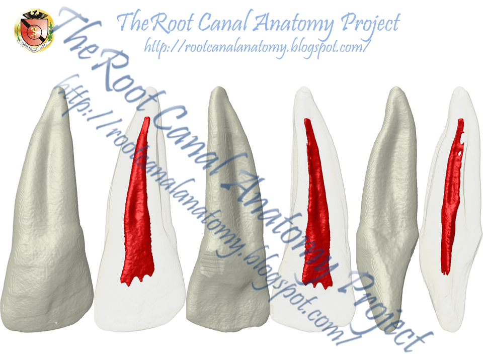
Morphology
The grooves also vary in depth. Variation in groove depth can make a communication possible with the pulp cavity, however, deep grooves with direct communications with the pulp are seldom reported. With increasing depth of the groove, the thickness of the root cementum increases. At a morphological level, RGs are characterized by reduced dentin thickness and an increased cement layer, with a simultaneous modification of the odontoblasts. At a histological level, irregular dentin cement junctions have typically been identified.

Etiology
The etiology of RGs is unknown. Similar to an invagination, this seems to be a peculiarity of tooth development accompanied by a further anomaly. Black was the first to describe the RG as a malfor-mation during embryo development in 1908. Atkinson surmised that the reason for its formation is that there is not enough space during tooth development in the maxilla, resulting in folding in the area of the Hertwig epithelial sheath. In the opinion of Goon et, this could also be an attempt at a root partition. According to recent studies, RGs may be caused by genetic changes. The predominance of the palato-gingival groove in the maxillary lateral incisors suggests the possibility that the groove results from an undesirable position of the lateral incisor during the period of maxilla growth. The tooth, although still a germ, becomes surrounded by the central incisor, canine, and first premolar that are in a more advanced phase of dental development. Mineralization of the crown of the maxillary lateral incisor starts later, compared with the others, making this germ, under these conditions, highly susceptible to folding.

Clinical remarks
Morphological defects in dental structure (e.g. dens invaginatus, talon cusp, and the palato-gingival groove) can be predisposing factors for the onset of inflammatory processes in the periodontal and/or pulp tissues. The funnel-like shape of the palato-gingival groove promotes the accumulation of difficult-to-remove plaque and calculus, at times making proper cleaning by the patient, or even by the dentist, nearly impossible. Most of the time RGs are clinically overlooked so that recurring clinical symptoms are often misdiagnosed because of the pathogenesis. As there is no epithelial closure, it is possible for microbes to settle in the groove. Depending on the morphology of the RG, localized periodontitis may develop, accompanied by pathosis. Periodontal pocket depths of more than 5 mm and increased dental mobility are typical findings. Moreover, in the case of deep RGs, a retrograde pulpitis may occur as a result of the so-called endodontic-periodontal lesion.
Treatment
A good prognosis for prolonged tooth preservation also depends on whether combined periodontal and endodontic therapy is necessary. Several different procedures have been proposed for successful correction of RG. Current clinical treatment can correspond to that for inflammatory periodontal diseases. In some cases the palato-gingival groove can be seen in periapical radiographs as a fine parapulpal radiolucent line. In most cases, odontoplasty was carried out in combination with regenerative therapy. In the presence of periodontal disease, the therapeutical options can consist of grinding and flattening the affected area of the tooth with the groove, with the placement of a physical barrier between the tooth and soft tissue flap. The intraosseous defect, if present, can be grafted with porous hydroxyapatite. Other reported treatment procedures are careful root planing and cleaning, filling of the groove with amalgam or calcium sulphate, and intentional replantation after root planing and the insertion of Emdogain. The inherent difficulties in treating the palato-gingival groove make its diagnosis complex for practitioners. In some cases, the tooth was extracted due to its high mobility or in cases of bruxism.
Prognosis
The prognosis of pulp diseases and/or periapical inflammation in the presence of a palato-gingival groove is not very favorable, and depends in part on the groove’s extension, the depth of the groove, and the relation of the groove to the pulp cavity.

Sources
Arnold M.Palato-radicular groove associated with a bi-rooted maxillary incisor: a case report. Endo 2007;1:205-213.
Pécora JD, Conrado CA, Zuccolotto WG, et al.: Root canal therapy of an anomalous maxillary central incisor: a case report. Endod Dent Traumatol 1993; 9: 260-262.
Pécora JD, da Cruz Filho AM: Study of the incidence of radicular grooves in maxillary incisors. Braz Dent J 1992; 3: 11-16.
Pecora JD, Sousa Neto MD, Santos TC, et al.: In vitro study of the incidence of radicular grooves in maxillary incisors. Braz Dent J 1991; 2: 69-73.
Simon JH, Dogan H, Ceresa LM, et al.: The radicular groove: its potential clinical significance. J Endod 2000; 26: 295-298.
No comments:
Post a Comment